

Cone Beam Imaging in Lakewood, CO
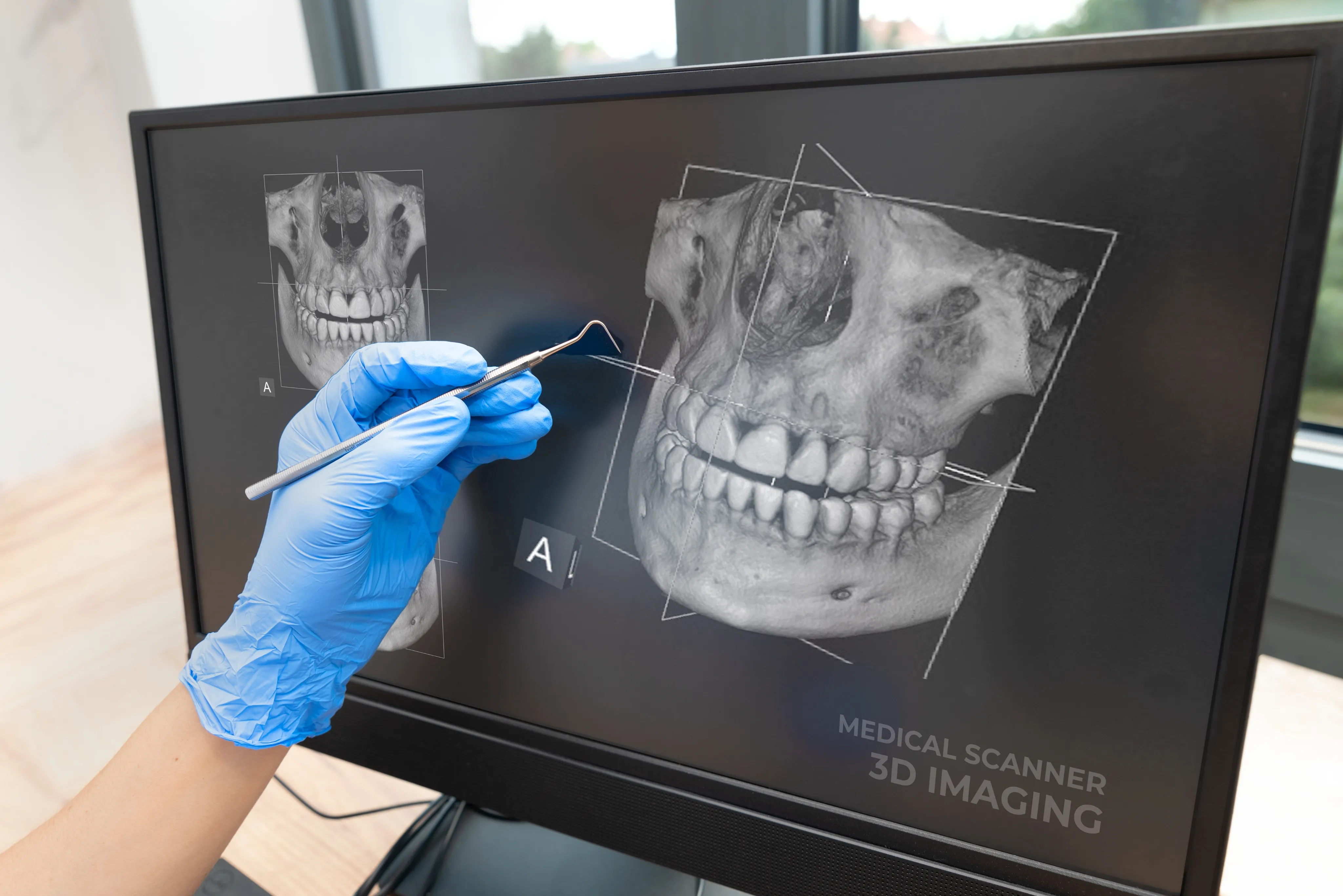
Cone beam imaging in Lakewood, Colorado, gives Dr. Dian Li a comprehensive 3D view of your oral structures. This advanced technology supports precise treatment planning for implants, extractions, and other complex procedures.
Cone Beam Imaging Explained
Cone beam imaging is an advanced type of dental X-ray that captures hundreds of images in a single rotation around your head. Specialized software assembles these images into a precise three-dimensional model of your mouth and jaw. Unlike a medical CT scan, a dental CBCT uses a cone-shaped beam and typically a lower radiation dose, tailored for dental and facial anatomy.
When It Is Recommended
- Dental implant planning to measure bone height, width, and density.
- Root canal evaluation to map canals, locate extra canals, and assess fractures.
- Wisdom teeth and impaction assessment, including nerve proximity.
- Temporomandibular joint (TMJ) and jaw joint analysis.
- Orthodontic planning and airway evaluation for bite and breathing concerns.
- Lesion, cyst, or pathology review when 2D X-rays are unclear.
Benefits of Cone Beam Imaging
- Enhanced diagnostic detail with a true 3D view of teeth, roots, nerves, and bone.
- More accurate treatment planning for implants, surgery, and endodontic care.
- Targeted, conservative care by seeing exactly where to treat.
- Fewer surprises during procedures with clearer anatomy mapping.
- Efficient single scan that often replaces multiple 2D X-rays.
- Lower dose than medical CT while providing far more information than traditional dental X-rays.
The Cone Beam Imaging Process
The CBCT scan is quick and comfortable. You will stand or sit while your head is gently positioned. The scanner rotates once around your head, usually in 10 to 20 seconds. It does not touch you, and you will not feel the X-rays. Remaining still ensures sharp images. After the scan, software reconstructs a detailed 3D model that can be rotated and measured from any angle.
Step-By-Step Overview
- Preparation: Remove glasses, earrings, and removable metal objects.
- Positioning: Bite lightly on a small guide or rest your chin as directed.
- Scanning: Keep still while the unit circles your head.
- Review: Our dentist evaluates the 3D images and discusses findings and options.
What to Expect
Most patients need no special preparation and can eat and drink as usual. If there is a chance of pregnancy, notify the team before any X-ray. A CBCT scan uses low-dose radiation compared with medical CT; the dose is higher than a single small dental X-ray but can be similar to or modestly higher than a panoramic X-ray, depending on the scan size. The scan is fast, quiet, and painless. Results are available right away, so our dentist can explain how cone beam imaging informs your diagnosis and care plan.
Patients often ask how CBCT compares with traditional dental X-rays. A CBCT provides dimensional measurements, cross-sectional views, and nerve mapping that flat images cannot offer. This level of detail supports more predictable outcomes, especially for implants, extractions near nerves, and complex root canals.
Frequently Asked Questions
Yes. A CBCT scan uses a lower radiation dose than a medical CT scan and is tailored for dental anatomy. The scan is brief, and exposure is kept as low as reasonably achievable while producing diagnostic-quality images.
The actual scan usually takes 10 to 20 seconds. Including positioning and review, the full process typically takes a few minutes.
No special preparation is needed. Remove glasses, earrings, and any removable metal objects. Eat and drink as usual. Notify the team if there is any chance of pregnancy.
A CBCT provides a three-dimensional view with cross-sectional slices, while a standard X-ray produces a flat, two-dimensional image. CBCT reveals depth, bone quality, and nerve positioning that flat images cannot show.
Coverage varies by plan and reason for the scan. Many plans cover CBCT when it is medically necessary for implant planning, impacted teeth, or pathology evaluation. Our team can help verify your benefits.
CBCT is not part of every visit. It is recommended when detailed 3D information is needed for specific diagnosis or treatment planning. Your dentist will explain why a scan is recommended and how it benefits your care.

Advanced Imaging for Better Outcomes
Contact Belmar Smiles at 303-935-3574 to learn how cone beam imaging can support your treatment plan.